Original Articles

Aquatic Nature. 30 June 2021. 57-69
https://doi.org/10.23135/an.2021.1.1.6

ABSTRACT


MAIN

  • 1. INTRODUCTION

  • 2. MATERIALS AND METHODS

  •   2.1 Sample collection, DNA extraction, and metagenome sequencing

  •   2.2 Analysis of assembly, gene prediction, functional annotations, and phylogeny

  • 3. RESULTS AND DISCUSSION

  •   3.1 Analysis of rhodolith metagenome sequences

  •   3.2 Carbon and nitrogen cycling in rhodolith holobiont

  • 4. CONCLUSION

1. INTRODUCTION

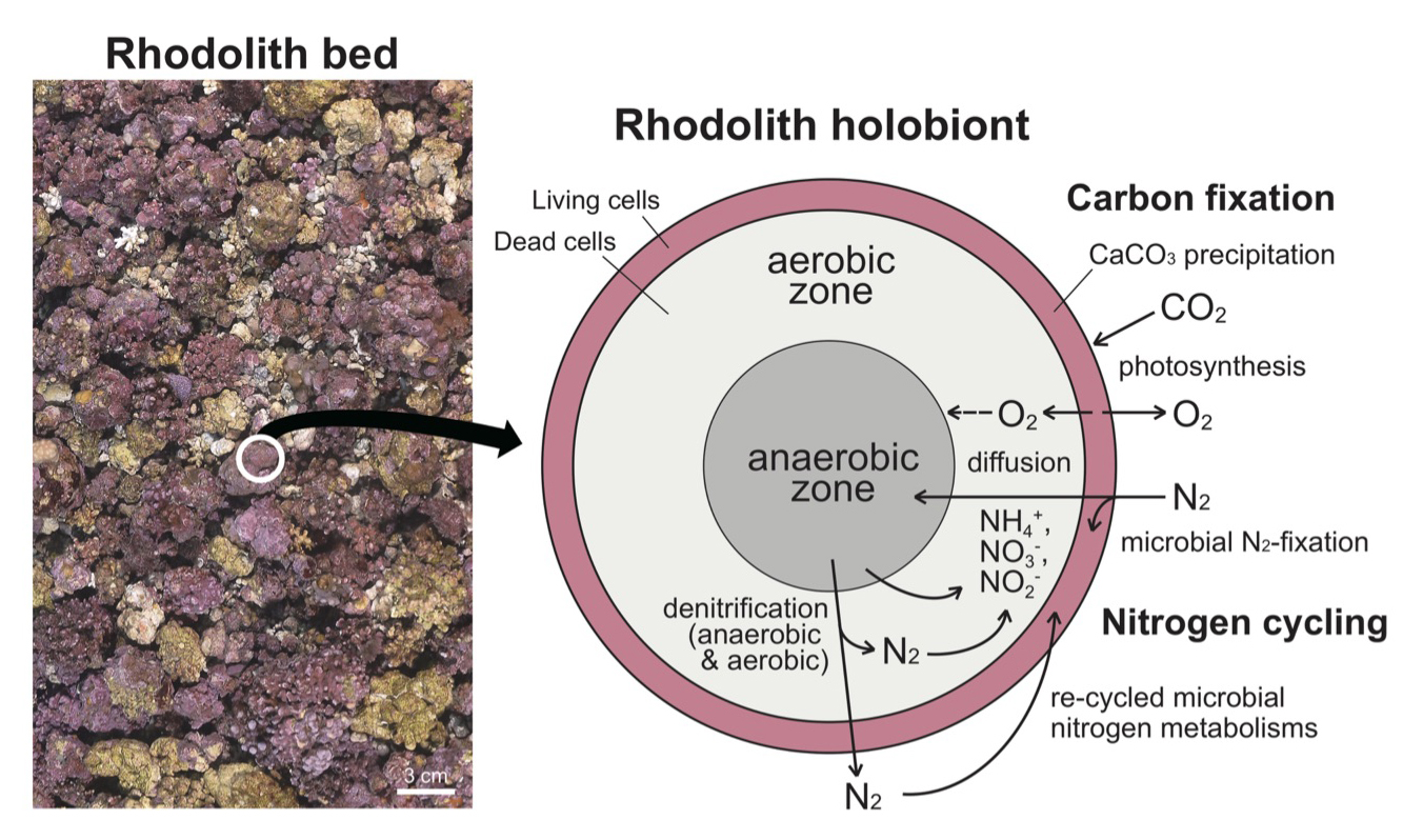
Rhodoliths are morphologically diverse and free-living non-geniculate coralline red algae (Rhodophyta), which are generally distributed in worldwide marine benthic habitats (Bosence 1983; Foster 2001; Fredericq et al. 2019; Jeong et al. 2019). Rhodolith beds are especially common from the low intertidal zone to the mesophotic zone (ca. ~150 m depth) along the continental shelf and near oceanic islands (Harris et al. 1996; Amado-Filho and Pereira-Filho 2012). An individual rhodolith is distinguished from crustose algae-coated pebble (McCoy and Kamenos 2015). The size and density of rhodoliths on the seafloor varies with water depth and latitude (Horta et al. 2016). Rhodoliths play an important ecological role by providing a microhabitat in a wide range of floral and faunal communities, e.g. kelp beds, corals, sponges, and herbivores, which are generally associated with biodiversity, abundance and food webs (Grall et al. 2006; Amado-Filho and Pereira-Filho 2012; Amado-Filho et al. 2017; Gabara 2020). For instance, a recent study showed numerous single-celled organisms including Prorocentrum lima (dinoflagellate) and Ochrosphaera verrucosa (haptophyte) are inhabiting within calcified cell lumens of rhodoliths using SEM, TEM and fluorescence micrographs (Krayesky-Self et al. 2017). Rhodolith beds also play crucial biogeochemical roles through photosynthesis and biomineralization of calcium carbonate (CaCO3) skeletons (Cabioch et al. 1999; Halfar et al. 2000; Williams et al. 2011; Kamenos and Law 2010; McCoy and Kamenos 2015). The depositions of calcified cell walls are constructed from dissolved inorganic carbon (DIC: aquatic CO2, HCO3-, and CO32-) and photosynthesis also responds to the elevated DIC in coralline algae (Gao et al. 1993; McCoy and Kamenos 2015). Carbonic anhydrases play crucial role in photosynthesis (Atkinson et al. 2016; Gee and Niyogi 2017; Razzak et al. 2019), and these enzymes are also involved in the calcifying mechanism of cnidarian coral skeletons (Bertucci et al. 2013). Rhodolith beds and crustose coralline algae are truly one of major contributors to carbonate deposition in the world’s oceans. These algae are hypothesized to be major contributors of the global ocean carbon biogeochemical cycling; however, biological interactions of rhodolith beds with global and local environmental changes are still unclear (Testa and Bosence 1999; Amado-Filho and Pereira-Filho 2012; Basso 2012; McCoy and Kamenos 2015; Horta et al. 2016; Teed et al. 2020).

Nitrogen metabolism is also involved in the carbon fixation mechanisms, and it is significantly important in algal growth (e.g., rhodolith beds) (Macler 1986; Huppe and Turpin 1994; Zhou et al. 2016; Schubert et al. 2019). However, usable nitrogen sources (e.g., ammonium, ammonia, nitrate) for eukaryotes are frequently acquired from microbial nitrogen fixers (N2-fixers); these diazotrophs include oxygenic/anoxygenic phototrophic, aerobic/anaerobic heterotrophic, bacteria, and archaea (Zhang et al. 2020). The epiphytic or symbiotic diazotrophs generate a nitrogen flux to rhodoliths and other macroalgal communities, and this nitrogen source leads to biological storage and cycling of both carbon and nitrogen within the marine trophic structure (Capone and Carpenter 1982; Phlips and Zeman 1990; Matson and Quenga 2005; Gabara 2020; Zehr and Capone 2020; Zhang et al. 2020). Rhodoliths and their microbial community consistently interact and form an ecological unit termed holobiont, which responds to environmental changes (Cavalcanti et al. 2018; van der Loos et al. 2019). The rhodolith holobionts include bacteria, archaea, virus, and diverse small eukaryotic organisms that were discovered by metagenomic approaches therefore rhodolith beds are ecologically important as hotspots of marine biodiversity, productivity, and resilience (Cavalcanti et al. 2013, 2014, 2018; Fredericq et al. 2019). However, biological interactions within the rhodolith holobiont are poorly studied.

To get more deeper understanding of rhodolith holobionts, we generated a metagenome sequencing data of a rhodolith using the whole-genome shotgun sequencing method, and we analyzed the species composition and its potential interactions. Our results reveal that the aerobic and anaerobic microbial community interact within the holobiont, playing an important role as the interactive channel for carbon and nitrogen cycling between biotic and abiotic environments around the rhodoliths.

2. MATERIALS AND METHODS

2.1 Sample collection, DNA extraction, and metagenome sequencing

The rhodolith samples were collected from Udo Island, Jeju, Korea (vicinity of 33°31'03.4"N 126°56'10.9"E; 2014.09.18; September 18, 2014), and placed in liquid nitrogen for several minutes. One of the frozen rhodolith samples was ground using a mortar and pestle, and total genomic DNA was extracted using the DNeasy Plant Mini Kit (Qiagen, Hilden, Germany). Whole-metagenome sequencing was conducted by Ion Torrent PGM platform with 400 bp-sized sequencing library kit (Thermo Fisher Scientific, San Francisco, California, USA). Raw reads (SRR12920572; https://www.ncbi.nlm.nih.gov/sra? linkname=bioproject_sra_all&from_uid=672827) and assembly (JADWZK000000000; https://www.ncbi.nlm.nih.gov/nuccore/ JADWZK000000000) of metagenome sequencing were uploaded to the NCBI SRA (Sequence Read Archive) database (BioProject PRJNA672827; https://www.ncbi.nlm.nih.gov/bioproject/PRJNA672827).

2.2 Analysis of assembly, gene prediction, functional annotations, and phylogeny

The sequenced metagenome raw reads were assembled by MEGAHIT assembler (Li et al. 2015). To classify the rhodolith sample, we analyzed 18S ribosomal DNA (rDNA) sequences using local BLASTn search (e-value cutoff = 1.e-10) with full length rDNA sequences of coralline algae (from Sporolithales, Rhodogorgonales, Hapalidiales, and Corallinales; Lee et al. 2018). The complete 18S rDNA sequences in the contig was analyzed by the web-based program RNAmmer 1.2 Server (Lagesen et al. 2007). Based on the assembly, protein sequences were analyzed by the Prodigal gene prediction software (Hyatt et al. 2010). Protein functions were analyzed by DIAMOND search (Buchfink et al. 2015) based on KEGG database (metabolic pathway analysis; http://www.genome.jp/tools/blast). The processes of assembly, gene prediction, and functional annotation were conducted by the SqueezeMeta pipeline (Tamames and Puente-Sánchez 2019). All BLAST and functional databases were downloaded using “download_databases.pl” script, which is involved in the SqueezeMeta pipeline (Tamames and Puente-Sánchez 2019). Taxonomic assignments of assembled contigs and analyzed genes in this study were defined by a representative taxa of BLAST/DIAMOND search results that analyzed by a fast LCA (lowest common ancestor; Luo et al. 2014) algorithm with nr database (Download: Aug. 2020) on the SqueezeMeta pipeline (Tamames and Puente-Sánchez 2019). The target gene was analyzed by BLASTp search (e-value cutoff = 1.e-05), and aligned with top 100 homologous sequences from the BLAST results using MAFFT v7.313 (default option: --auto; Yamada et al. 2016). Phylogenetic analysis was done using the maximum likelihood (ML) method with 1,000 bootstrap replications, and the best-fit evolutionary model was selected by default selection option (IQ-tree v1.6.12; Nguyen et al. 2015).

3. RESULTS AND DISCUSSION

3.1 Analysis of rhodolith metagenome sequences

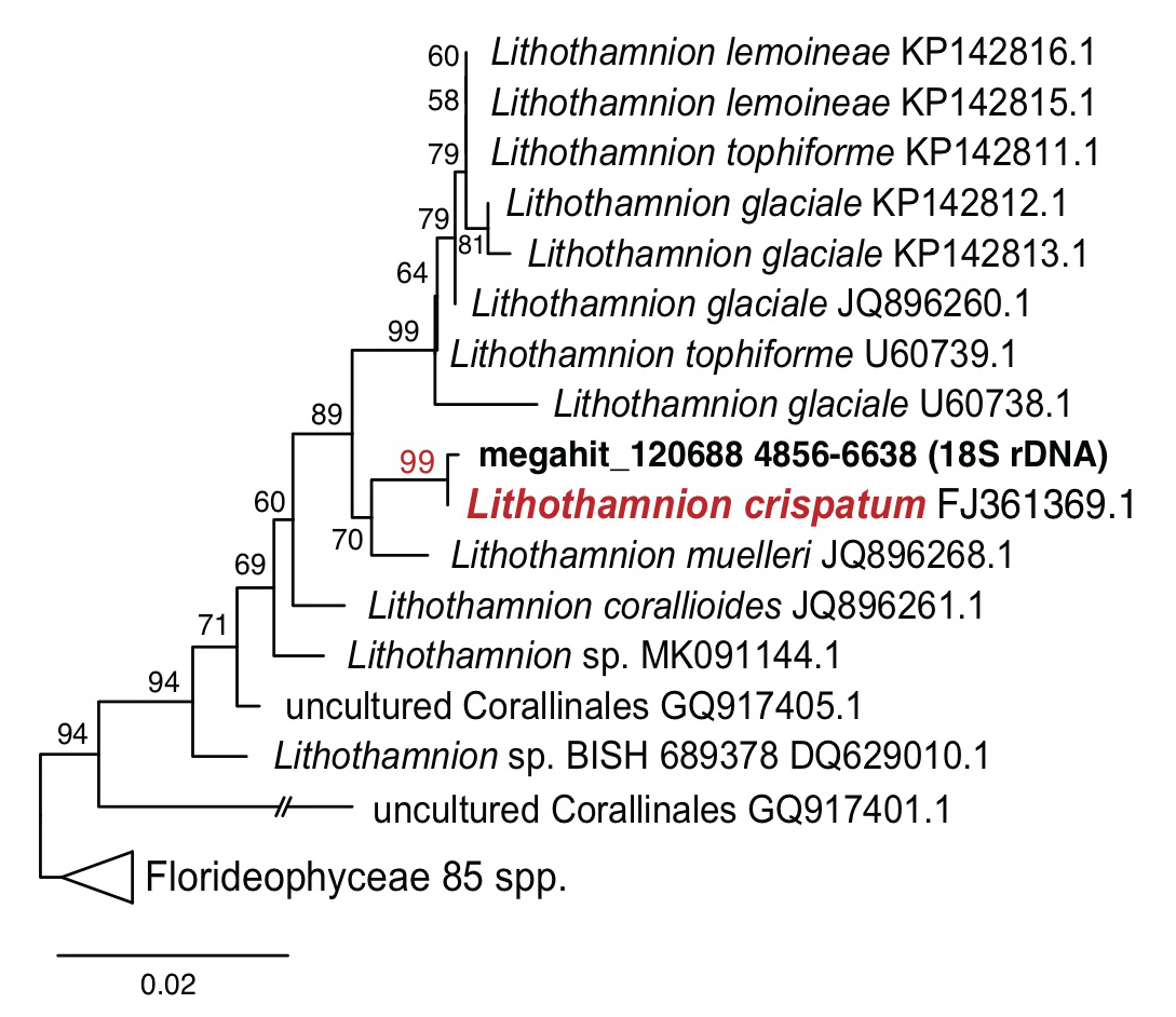
The generated rhodolith metagenome sequencing data (1.3 Gbp, the Ion Torrent PGM platform; Thermo Fisher Scientific, San Francisco, California) were assembled (210 Mbp; 376,038 contigs; N50=581 bp) by MEGAHIT assembler (Li et al. 2015). Based on the assembly, a total of 395,055 proteins was analyzed by the Prodigal gene prediction software (Hyatt et al. 2010). From the assembly, we found 18S ribosomal DNA (rDNA) sequences in contig ‘megahit_120688’, which is 1,783 bp in length, and this shows 99.9% sequence similarity and a strong monophyletic relationship with Lithothamnion crispatum (FJ361369.1; Fig. 1). A non-geniculate coralline alga, L. crispatum is a cosmopolitan rhodolith forming alga that is known to play an important role in biogenic calcium carbonate deposition (Basso et al. 2011; Amado-Filho et al. 2012; de Carvalho et al. 2017).

https://static.apub.kr/journalsite/sites/ksp/2021-001-01/N0310010106/images/ksp_01_01_06_F1.jpg
Fig. 1

Phylogenetic tree of the rhodolith 18S rDNA sequences with top 100 homologous sequences from BLASTn search (e-value cutoff = 1.e-10).

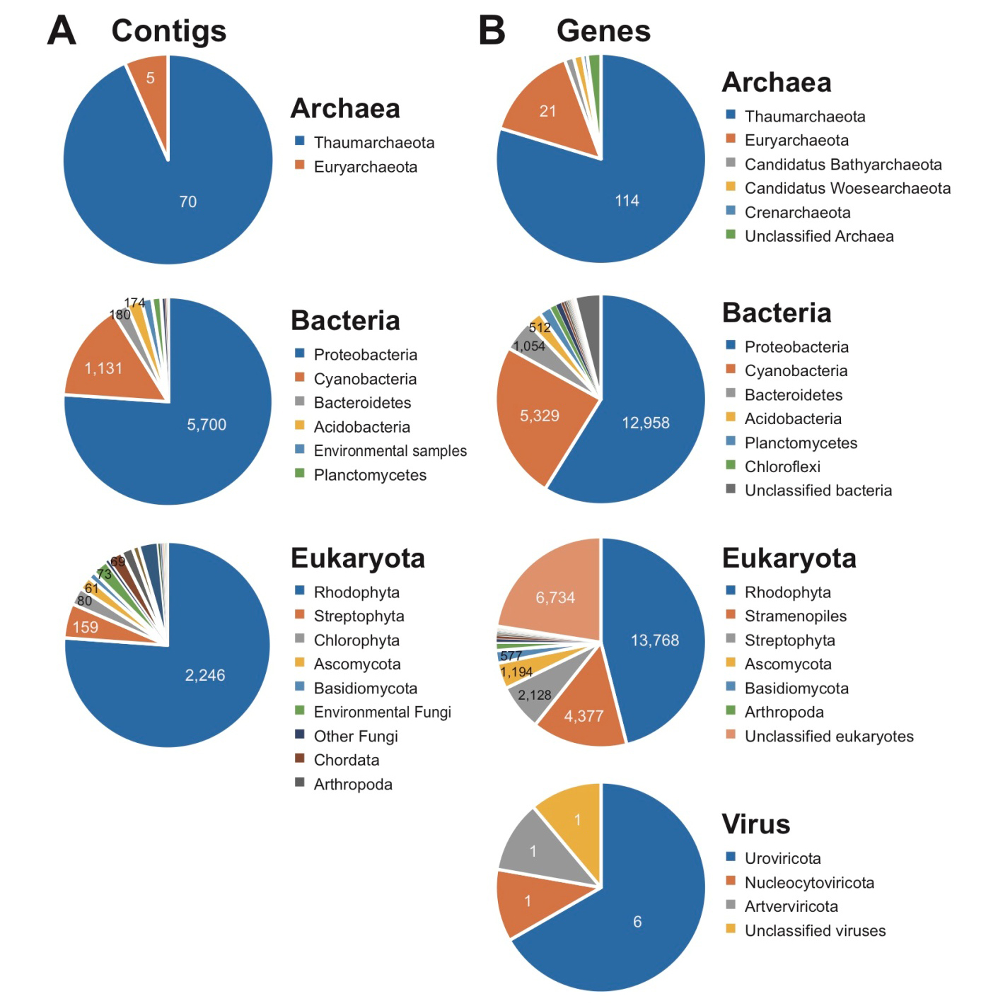
BLASTn analysis (e-value cutoff = 1.e-10) was done using 376,052 assembled contigs with nt database (Download: Oct. 2019), and a total of 10,541 contigs shows BLAST top hits to Archaea (75), Bacteria (7,493), and Eukaryota (2,950), Virus (1), and unclassified sequences (22), including Thaumarchaeota (70; Archaea), Proteobacteria (5,700; Bacteria), and Rhodophyta (2,246; Eukaryota), respectively (Fig. 2A). In eukaryotic contigs, fragmented plastid (1,127 contigs) and mitochondrial (702 contigs) genomes were found representing diverse taxonomic groups: identified plastid genome fragments from Rhodophyta (1,039), Chlorophyta (41), Stramenopiles (32), Streptophyta (11), Cryptophyta, (2), Euglenophyta (1), unclassified plastid (1) and mitochondrial genome fragments from Rhodophyta (661), Metazoa (16), Stramenopiles (9), Streptophyta (6), Fungi (6), Jakobida (2), Chlorophyta (1), Amoebozoa (1). Interestingly, BLAST top match sequences of fragmented 16 plastid contigs in our metagenome assembly belong to the chlorophyte algae Ostreobium sp. (KY819067, KY766991, KY766993, KY766995, and KY766996) and O. quekettii (LT593849); Ostreobium is the most abundant endolithic algal genus in marine environments, especially in cnidarian coral holobionts (Marcelino and Verbruggen 2016; Verbruggen et al. 2017; Massé et al. 2018). Ostreobium is also known to be a major species for biogenic dissolution of carbonates from coral reefs to the open ocean, which may have a significant impact on the calcium carbonate budget of coral reefs (Aline 2008; Grange et al. 2015). Our results suggest that Ostreobium may also contribute to the bioerosion of carbonates in rhodolith holobionts.

A total of 395,055 genes was analyzed in the metagenome assembly, and 55,399 genes were annotated based on taxonomic assignments of the analyzed genes (Supplementary Table 1; Fig. 2B). Although the metagenome data cannot represent quantification of species abundance, the taxonomic composition of the genes includes Archaea (143), Bacteria (22,016), and Eukaryota (29,896), Viruses (9), and unclassified taxa (3,335), and their major taxa are consistent with the results using contigs as follows: Thaumarchaeota (114; Archaea), Proteobacteria (12,958; Bacteria), and Rhodophyta (13,768; Eukaryota) (Fig. 2B). However, the results of taxonomic annotations using the genes show a more diverse taxonomic composition than those for the contigs (Fig. 2A and 2B). Among Rhodophyta genes, most of them (94.2%) were identified as the Florideophyceae that includes the subclass Corallinophycidae (i.e., coralline algae). The Bangiophyceae genes (2.3%) are present, indicating filamentous and foliose seaweeds (Blouin et al. 2011; Kucera and Saunders 2012; Koh and Kim 2018). Our rhodolith could include unicellular red algae (i.e., class Porphyridiophyceae and Cyanidiophyceae; genes < 2%, respectively among red algal genes). Because only several red algal genomes are available in the NCBI database, species-level compositional studies require additional red algal genomes. The Streptophyta genes (Supplementary Table 1) are regarded as seagrass genes but some of genes could be other algal genes because many genes in primary endosymbiosis groups show monophyletic relationships with other sister algal lineages (Chan et al. 2011). Other, diverse photosynthetic algal lineages are also present; brown algae (229 genes), diatoms (206 genes), green algae (51 genes), dinoflagellates (13 genes), haptophytes (8 genes), and apicomplexans (8 genes). This result corresponds well with natural habitats of Ecklonia cava and Undaria peterseniana (Hwang et al. 2010). Several dinoflagellates and haptophytes have a benthic stage (e.g., cysts) that is endolithically associated with rhodolith holobionts (Krayesky-Self et al. 2017). We found 13 genes of Scleractinia group (stony corals), which provide potential evidence for physical interactions between calcifying coralline algae and cnidarian corals. Cnidarian corals frequently contain symbionts (e.g., Symbiodinium) or parasites (e.g., apicomplexan-related lineage) (Janouškovec et al. 2012, 2013; González-Pech et al. 2019). Regardless of whether the apicomplexan genes in our metagenome data are directly associated with the rhodolith cells or the coral cells, it is evident that complex communities form the holobiont. Interestingly, oomycetes (water molds; non-photosynthetic Stramenopiles; 3,985 genes) shows relatively larger proportion in the rhodolith metagenome data. The oomycetes are indicated as fungi-like pathogens or parasites in plant hosts, which are physiologically related each other (Birch et al. 2006; Thines and Kamoun 2010; Richards et al. 2011; Pais et al. 2013). We suggest that rhodoliths also have a host-parasite interaction.

https://static.apub.kr/journalsite/sites/ksp/2021-001-01/N0310010106/images/ksp_01_01_06_F2.jpg
Fig. 2

Taxonomic assignments of metagenome analysis results. A. BLASTn search results of assembled contigs. B. Protein-coding genes analyzed by DIAMOND search results.

3.2 Carbon and nitrogen cycling in rhodolith holobiont

We focused on carbon and nitrogen cycle genes from the functional annotations of genes. A total of 25 carbonic anhydrases (CAs) was found in the rhodolith holobiont: 20 are regarded as bacterial, four as eukaryotic, and one with an unknown origin (Supplementary Table 2). The CA genes have an important role of capturing CO2, which is used in crucial cellular processes (e.g., photosynthesis) called carbon concentrating mechanism (Badger and Price 1994; Supuran and Capasso 2017; Wang et al. 2020). Diverse photosynthetic organisms in the rhodolith holobiont are involved in carbon cycling through the photosynthetic carbon fixation. Especially, coralline algae (i.e., rhodolith) may actively use the mechanism to deposit carbon sources not only through their plastid (photosynthesis) but also calcified cell walls (calcification) based on the dissolved inorganic carbon sources such as another calcifying organism, cnidarian corals (Gao et al. 1993; Bertucci et al. 2013; McCoy and Kamenos 2015; Wang et al. 2020).

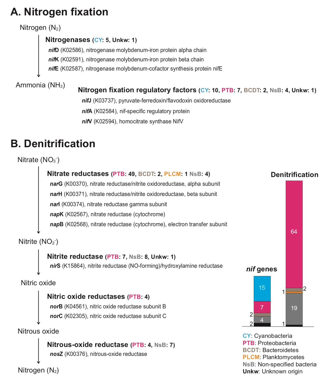
We found the following diverse genes involving nitrogen fixation (nif): nifD (K02586) and nifK (K02591) for nitrogenase molybdenum-iron protein function, nifE (K02587) for nitrogenase molybdenum-cofactor synthesis protein, nifJ (K03737) for pyruvate-ferredoxin/flavodoxin oxidoreductase, nifA (K02584) for nif-specific regulatory protein, nifV (K02594) for homocitrate synthase protein, and nif11 domain (pfam07862) containing protein (Supplementary Table 3). Among them, nifD, nifK, and nifE genes have core function (nitrogenase) during nitrogen fixation, directly converting nitrogen gas (N2) to ammonia (NH3) (Fani et al. 2000). Most of nitrogenase genes and their regulatory factors in the rhodolith holobiont were cyanobacterial genes (Fig. 3A, and Supplementary Table 3). In marine environments, biological nitrogen fixation of microbial communities is an important factor to supply usable nitrogen sources as key nutrients to algal cells through assimilation, and the nitrogen fixers are present in diverse algal holobionts (Cheng 2008; Barott et al. 2011; Sohm et al. 2011; de Oliveira et al. 2012; Egan et al. 2013). Several non-cyanobacterial (e.g., proteobacterial) nif regulatory factors were also present in the rhodolith holobiont (Fig. 3A, and Supplementary Table 3), and these are possibly indicated as non-cyanobacterial nitrogen fixers (Riemann et al. 2010; Farnelid et al. 2011; Sohm et al. 2011; Delmont et al. 2018). Based on these findings, we suggest that microbial nitrogen fixers in rhodolith populations contribute to nitrogen cycling. Interestingly, there are many different kinds of denitrification genes that include nitrate reductases (narG, narH, narI, napK, and napB), nitrite reductase (nirS), nitric oxide reductases (norB, and norC), and nitrous-oxide reductase (nosZ) (Fig. 3B, and Supplementary Table 4). Most denitrification genes are identified as proteobacterial genes (i.e., alphaproteobacterial, gammaproteobacterial, deltaproteobacterial, and unidentified proteobacterial genes, see Supplementary Table 4). Proteobacteria groups are abundant in diverse environments, and play an important ecological role in carbon, sulfur, and nitrogen cycles. Among the proteobacterial groups, deltaproteobacteria include diverse anaerobic families (Greene 2014; Kuever 2014a, 2014b). Several strict anaerobic deltaproteobacterial families including Desulfobacteraceae, Desulfobulbaceae, and Desulfuromonadaceae (strict but some tolerance to oxygen) are also present in the rhodolith holobiont, and they have nitrogen fixation and denitrification functions (Supplementary Table 3, and 4; Greene 2014; Kuever 2014a, 2014b). Indeed, it was reported that anaerobic sediments were preserved in rhodolith beds (Adey et al. 2015). We suggest the rhodolith holobiont generally has an anaerobic zone too, and their anaerobic bacterial community contributes the nitrogen cycling along with other aerobic/aerotolerant populations. The diffusion of oxygen in the particle microenvironments can be limited by the diffusive boundary layer more than nitrate, and anaerobic nitrogen metabolisms can occur in the central area of the microenvironment (Bianchi et al. 2018). Therefore, the re-cycled nitrogen may occur between the nitrogen fixation and denitrification organisms within the rhodolith holobiont (i.e., the inside of crustose algae-coated pebble including deposition of calcium carbonate). However, actual measurements of carbon and nitrogen flows in rhodolith are still required because our metagenomics approach can only reveal functional potential and cannot distinguish dormant microorganisms (Lennon and Jones 2011).

https://static.apub.kr/journalsite/sites/ksp/2021-001-01/N0310010106/images/ksp_01_01_06_F3.jpg
Fig. 3

Nitrogen fixation and denitrification pathways in metagenome results. A. Nitrogen fixation and their regulatory factor genes with their phylogenetic positions. B. Denitrification genes with their phylogenetic positions.

4. CONCLUSION

Rhodoliths (coralline algae) may support crucially for carbon cycling through calcium carbonate precipitations in their cell walls and photosynthesis, even though the cellular activities are only present in living cells of surface area (Fig. 4). Cyanobacterial communities also make photosynthetic activities in the photic surface of rhodoliths, and they conduct nitrogen fixation mechanisms (Supplementary Table 2 and 3), which could use in cell growth of rhodolith as well as other epiphytic algal community. The active interaction between cyanobacteria and rhodolith algae could accelerate carbon cycling. On the other hand, dead cells were filled inside of rhodolith (Fig. 4). These calcified dead cells within rhodolith serve habitats for diverse microbials communities as seedbanks and temporal reservoirs (Krayesky-Self et al. 2017; Fredericq et al. 2019). Because we found strict anaerobic microbial community (i.e., Desulfobacteraceae, Desulfobulbaceae) from our metagenome data as well as deltaproteobacterial species from rhodolith holobionts in a previous metagenome study (Cavalcanti et al. 2018), it is likely that the rhodolith holobiont comprises of aerobic and anaerobic zones as a functionally enclosed biosphere (Fig. 4). Interestingly, both aerobic and anaerobic denitrifications occur in rhodolith holobiont, and these re-cycled nitrogen sources may be used in nitrogen fixation again within the holobiont. The internal microbial interactions within the rhodolith holobiont may have advantages to maintain their biodiversity and respond directly to local environmental changes as self-sustaining reservoirs for marine carbon and nitrogen cycling. More metagenome data from rhodoliths could clarify the heterogeneity of microhabitats (e.g., surface and interior parts; Kim et al. 2021), and also reveal the major functions of the community structures (Amado-Filho and Pereira-Filho 2012; Amado-Filho et al. 2017; Cavalcanti et al. 2018).

https://static.apub.kr/journalsite/sites/ksp/2021-001-01/N0310010106/images/ksp_01_01_06_F4.jpg
Fig. 4

Rhodolith bed from natural habitats (left) and the potential model of environmental interactions in rhodolith holobionts (right) for marine carbon and nitrogen cycling.

Acknowledgements

The authors thank to Sung Min Boo and Yong Deok Ko for collection of rhodolith samples, and Jeong Ha Kim for the image of rhodolith bed, which was taken in March of 2019 from the sampling site. We thank to Robert A. Andersen for valuable comments. This research was supported by the Collaborative Genome Program of the Korea Institute of Marine Science and Technology Promotion (KIMST) funded by the Ministry of Oceans and Fisheries (MOF) (20180430) and the National Research Foundation of Korea (NRF-2017R1A2B3001923 and 2020R1C1C1010193).

References

1
Adey WH and Macintyre IG. 1973. Crustose coralline algae: a re-evaluation in the geological sciences. Geol. Soc. Am. Bull. 84, 883-904. 10.1130/0016-7606(1973)84<883:CCAARI>2.0.CO;2
2
Adey W, Halfar J, Humphreys A, Suskiewicz T, Belanger D, Gagnon P and Fox M. 2015. Subarctic rhodolith beds promote longevity of crustose coralline algal buildups and their climate archiving potential. Palaios 30, 281-293. 10.2110/palo.2014.075
3
Aline T. 2008. Dissolution of dead corals by euendolithic microorganisms across the Northern Great Barrier Reef (Australia). Microb. Ecol. 55, 569-580. 10.1007/s00248-007-9302-617690835
4
Amado-Filho GM and Pereira-Filho GH. 2012. Rhodolith beds in Brazil: a new potential habitat for marine bioprospection. Rev. Bras. Farmacogn. 22, 782-788. 10.1590/S0102-695X2012005000066
5
Amado-Filho GM, Moura RL, Bastos AC, Salgado LT, Sumida PY, Guth AZ, Francini-Filho RB, Pereira-Filho GH, Abrantes DP, Brasileiro PS, Bahia RG, Leal RN, Kaufman L, Kleypas JA, Farina M and Thompson FL. 2012. Rhodolith beds are major CaCO3 bio-factories in the Tropical South West Atlantic. PLoS ONE 7, e35171. 10.1371/journal.pone.003517122536356PMC3335062
6
Amado-Filho GM, Bahia RG, Pereira-Filho GH and Longo LL. 2017. South Atlantic rhodolith beds: latitudinal distribution, species composition, structure and ecosystem functions, threats and conservation status. In: Rhodolith/maërl beds: a global perspective. Riosmena-Rodríguez R, Nelson W and Aguirre J, eds, Springer, Cham, 299-317. 10.1007/978-3-319-29315-8_12
7
Atkinson N, Feike D, Mackinder LCM, Meyer MT, Griffiths H, Jonikas MC, Smith AM and McCormick AJ. 2016. Introducing an algal carbon-concentrating mechanism into higher plants: location and incorporation of key components. Plant Biotechnol. J. 14, 1302-1315. 10.1111/pbi.1249726538195PMC5102585
8
Badger MR and Price GD. 1994. The role of carbonic anhydrase in photosynthesis. Annu. Rev. Plant Physiol. 45, 369-392. 10.1146/annurev.pp.45.060194.002101
9
Barott KL, Rodriguez-Brito B, Janouškovec J, Marhaver KL, Smith JE, Keeling P and Rohwer FL. 2011. Microbial diversity associated with four functional groups of benthic reef algae and the reef-building coral Montastraea annularis. Environ. Microbiol. 13, 1192-1204. 10.1111/j.1462-2920.2010.02419.x21272183
10
Basso D, Rodondi G and Bressan G. 2011. A re-description of Lithothamnion crispatum and the status of Lithothamnion superpositum (Rhodophyta, Corallinales). Phycologia 50, 144-155. 10.2216/10-20.1
11
Basso D. 2012. Carbonate production by calcareous red algae and global change. Geodiversitas 34, 13-34. 10.5252/g2012n1a2
12
Bertucci A, Moya A, Tambutté S, Allemand D, Supuran CT and Zoccola D. 2013. Carbonic anhydrases in anthozoan corals - a review. Bioorg. Med. Chem. 21, 1437-1450. 10.1016/j.bmc.2012.10.02423199478
13
Bianchi D, Weber TS, Kiko R and Deutsch C. 2018. Global niche of marine anaerobic metabolisms expanded by particle microenvironments. Nat. Geosci. 11, 263-268. 10.1038/s41561-018-0081-0
14
Birch PRJ, Rehmany AP, Pritchard L, Kamoun S and Beynon JL. 2006. Trafficking arms: oomycete effectors enter host plant cells. J. Mol. Evol. 14, 8-11. 10.1016/j.tim.2005.11.00716356717
15
Blouin NA, Brodie JA, Grossman AC, Xu P and Brawley SH. 2011. Porphyra: a marine crop shaped by stress. Trends Plant Sci. 16, 1360-1385. 10.1016/j.tplants.2010.10.00421067966
16
Bosence DWJ. 1983. Coralline algal reef frameworks. J. Geol. Soc. 140, 365-376. 10.1144/gsjgs.140.3.0365
17
Buchfink B, Xie C and Huson DH. 2015. Fast and sensitive protein alignment using DIAMOND. Nat. Methods 12, 59-60. 10.1038/nmeth.317625402007
18
Cabioch G, Montaggioni LF, Faure G and Ribaud-Lauernti A. 1999. Reef coralgal assemblages as recorders of paleobathymetry and sea level changes in the Indo-Pacific province. Quat. Sci. Rev. 18, 1681-1695. 10.1016/S0277-3791(99)00014-1
19
Capone DG and Carpenter EJ. 1982. Nitrogen fixation in the marine environment. Science 217, 1140-1142. 10.1126/science.217.4565.114017740970
20
Cavalcanti GS, Gregoracci GB, Longo LDL, Bastos AC, Ferreira CM, Francini-Filho RB, Parahos R, Ghisolfi RD, Krüger R, Güth AZ, Sumida PYG, Bruce T, Maia-Neto O, Santos EO, Iida T, Moura RL, Amado-Filho GM and Thompson FL. 2013. Sinkhole-like structures as bioproductivity hotspots in the Abrolhos Bank. Cont. Shelf Res. 70, 126-134. 10.1016/j.csr.2013.05.011
21
Cavalcanti GS, Gregoracci GB, Santos EOD, Silveira CB, Meirelles PM, Longo L, Gotoh K, Nakamura S, Iida T, Sawabe T, Rezende CE, Francini-Filho RB, Moura RL, Amado-Filho GM and Thompson FL. 2014. Physiologic and metagenomic attributes of the rhodoliths forming the largest CaCO3 bed in the South Atlantic Ocean. ISME J. 8, 52-62. 10.1038/ismej.2013.13323985749PMC3869012
22
Cavalcanti GS, Shukla P, Morris M, Ribeiro B, Foley M, Doane MP, Thompson CC, Edwards MS, Dinsdale EA and Thompson FL. 2018. Rhodoliths holobionts in a changing ocean: host-microbes interactions mediate coralline algae resilience under ocean acidification. BMC Genomics 19, 701. 10.1186/s12864-018-5064-430249182PMC6154897
23
Chan CX, Yang EC, Banerjee T, Yoon HS, Martone PT, Estevez JM and Bhattacharya D. 2011. Red and green algal monophyly and extensive gene sharing found in a rich repertoire of red algal genes. Curr. Biol. 21, 328-333. 10.1016/j.cub.2011.01.03721315598
24
Cheng Q. 2008. Perspectives in biological nitrogen fixation research. J. Integr. Plant Biol. 50, 786-798. 10.1111/j.1744-7909.2008.00700.x18713389
25
de Carvalho RT, Salgado LT, Amado-Filho GM, Leal RN, Werckmann J, Rossi AL, Campos APC, Karez CS and Farina M. 2017. Biomineralization of calcium carbonate in the cell wall of Lithothamnion crispatum (Hapalidiales, Rhodophyta): correlation between the organic matrix and the mineral phase. J. Phycol. 53, 642-651. 10.1111/jpy.1252628258584
26
de Oliveira LS, Gregoracci GB, Silva GGZ, Salgado LT, Amado-Filho G, Alves-Ferreira M, Pereira RC and Thompson FL. 2012. Transcriptomic analysis of the red seaweed Laurencia dendroidea (Florideophyceae, Rhodophyta) and its microbiome. BMC Genomics 13, 487. 10.1186/1471-2164-13-48722985125PMC3534612
27
Delmont TO, Quince C, Shaiber A, Esen ÖC, Lee ST, Rappé MS, McLellan SL, Lücker S and Eren AM. 2018. Nitrogen-fixing populations of Planctomycetes and Proteobacteria are abundant in surface ocean metagenomes. Nat. Microbiol. 3, 804-813. 10.1038/s41564-018-0176-929891866PMC6792437
28
Egan S, Harder T, Burke C, Steinberg P, Kjelleberg S and Thomas T. 2013. The seaweed holobiont: understanding seaweed-bacteria interactions. FEMS Microbiol. Rev. 37, 462-476. 10.1111/1574-6976.1201123157386
29
Fani R, Gallo R and Liò P. 2000. Molecular evolution of nitrogen fixation: the evolutionary history of the nifD, nifK, nifE, and nifN genes. J. Mol. Evol. 51, 1-11. 10.1007/s00239001006110903367
30
Farnelid H, Andersson AF, Bertilsson S, Al-Soud WA, Hansen LH, Sørensen S, Steward GF, Hagström Å and Riemann L. 2011. Nitrogenase gene amplicons from global marine surface waters are dominated by genes of non-cyanobacteria. PLoS One 6, e19223. 10.1371/journal.pone.001922321559425PMC3084785
31
Fredericq S, Krayesky-Self S, Sauvage T, Richards J, Kittle R, Arakaki N, Hickerson E and Schmidt WE. 2019. The critical importance of rhodoliths in the life cycle completion of both macro- and microalgae, and as holobionts for the establishment and maintenance of marine biodiversity. Front. Mar. Sci. 5, 502. 10.3389/fmars.2018.00502
32
Gabara SS. 2020. Trophic structure and potential carbon and nitrogen flow of a rhodolith bed at Santa Catalina Island inferred from stable isotopes. Mar. Biol. 167, 30. 10.1007/s00227-019-3635-9
33
Gao K, Aruga Y, Asada K, Ishihara T, Akano T and Kiyohara M. 1993. Calcification in the articulated coralline alga Corallina pilulifera, with special reference to the effect of elevated CO2 concentration. Mar. Biol. 117, 129-132. 10.1007/BF00346434
34
Gee CW and Niyogi KK. 2017. The carbonic anhydrase CAH1 is an essential component of the carbon-concentrating mechanism in Nannochloropsis oceanica. Proc. Natl. Acad. Sci. U.S.A. 114, 4537-4542. 10.1073/pnas.170013911428396394PMC5410810
35
Grall J, Loc'h FL, Guyonnet B and Riera P. 2006. Community structure and food web based on stable isotopes (δ15N and δ13C) analysis of a North Eastern Atlantic maerl bed. J. Exp. Mar. Biol. Ecol. 338, 1-15. 10.1016/j.jembe.2006.06.013
36
Grange JS, Rybarczyk H and Tribollet A. 2015. The three steps of the carbonate biogenic dissolution process by microborers in coral reefs (New Caledonia). Environ. Sci. Pollut. Res. 22, 13625-13637. 10.1007/s11356-014-4069-z25592911
37
Greene AC. 2014. The Family Desulfuromonadaceae. In: The Prokaryotes Deltaproteobacteria and Epsilonproteobacteria. Rosenberg E, DeLong EF, Lory S, Stackebrandt E and Thompson F, eds, Springer-Verlag, Berlin Heidelberg, 143-155.
38
Halfar J, Zack T, Kronz A and Zachos JC. 2000. Growth and high-resolution paleoenvironmental signals of rhodoliths (coralline red algae): a new biogenic archive. J. Geophys. Res. 105, 22107-22116. 10.1029/1999JC000128
39
Harris PT, Tsuji Y, Marshall JF, Davies PJ, Honda N and Matsuda H. 1996. Sand ans rhodolith-gravel entrainment on the mid- to outer-shelf under a western boundary current: Fraser Island continental shelf, eastern Australia. Mar. Geol. 129, 313-330. 10.1016/0025-3227(96)83350-0
40
Horta PA, Riul P, Amado-Filho GM, Gurgel CFD, Berchez F, Nunes JMDC, Scherner F, Pereira S, Lotufo T, Peres L, Sissini M, Bastos EO, Rosa J, Munoz P, Martins C, Gouvêa L, Carvalho V, Bergstrom E, Schubert N, Bahia RG, Rodrigues AC, Rörig L, Barufi JB and Figueiredo M. 2016. Rhodoliths in Brazil: current knowledge and potential impacts of climate change. Braz. J. Oceanogr. 64, 117-136. 10.1590/S1679-875920160870064sp2
41
Huppe HC and Turpin DH. 1994. Integration of carbon and nitrogen metabolism in plan and algal cells. Annu. Rev. Plant Biol. 45, 577-607. 10.1146/annurev.pp.45.060194.003045
42
Hwang EK, Gong YG and Park CS. 2010. Ecological characteristics of the endangered brown alga, Undariopsis peterseniana (Kjellman) Miyabe et Okamura, at Jeju Island, Korea: growth and maturation. Korean J. Fish. Aquat. Sci. 43, 63-68. 10.5657/kfas.2010.43.1.063
43
Hyatt D, Chen GL, LoCascio PF, Land ML, Larimer FW and Hauser LJ. 2010. Prodigal: prokaryotic gene recognition and translation initiation site identification. BMC Bionfiormatics 11, 119. 10.1186/1471-2105-11-11920211023PMC2848648
44
Janouškovec J, Horák A, Barott KL, Rohwer FL and Keeling PJ. 2012. Global analysis of platid diversity reveals apicomplexan-related lineages in coral reefs. Curr. Biol. 22, R518-R519. 10.1016/j.cub.2012.04.04722789997
45
Janouškovec J, Horák A, Barott KL, Rohwer FL and Keeling PJ. 2013. Environmental distribution of coral-associated relatives of apicomplexan parasites. ISME J. 7, 444-447. 10.1038/ismej.2012.12923151646PMC3554414
46
Jeong JB, Kim SY, Seo YK, Kim JK, Shin J and Woo KS. 2020. Influence of submarine topography and associated sedimentary processes on the distribution of live and dead rhodoliths near Udo Island, Korea. Geo-Mar. Lett. 40, 35-51. 10.1007/s00367-019-00623-w
47
Kamenos NA and Law A. 2010. Temperature controls on coralline algal skeletal growth. J. Phycol. 46, 331-335. 10.1111/j.1529-8817.2009.00780.x
48
Kim JH, Steller DL and Edwards MS. 2021. Variation in photosynthetic performance relative to thallus microhabitat heterogeneity in Lithothamnion australe (Rhodophyta, Corallinales) rhodoliths. J. Phycol. 57, 234-244. 10.1111/jpy.1308033020935
49
Koh YH and Kim MS. 2018. DNA barcoding reveals cryptic diversity of economic red algae, Pyropia (Bangiales, Rhodophyta): description of novel species from Korea. J. Appl. Phycol. 30, 3425-3434. 10.1007/s10811-018-1529-8
50
Krayesky-Self S, Schmidt WE, Phung D, Henry C, Sauvage T, Camacho O, Felgenhauer BE, Fredericq S. 2017. Eukaryotic life inhabits rhodolith-forming coralline algae (Haplidiales, Rhodophyta), remarkable marine benthic microhabitats. Sci. Rep. 7, 45850. 10.1038/srep4585028368049PMC5377461
51
Kucera H and Saunders GW. 2012. A survey of Bangiales (Rhodophyta) based on multiple molecular markers reveals cryptic diversity. J. Phycol. 48, 869-882. 10.1111/j.1529-8817.2012.01193.x27008998
52
Kuever J. 2014a. The Family Desulfobacteraceae. In: The Prokaryotes Deltaproteobacteria and Epsilonproteobacteria. Rosenberg E, DeLong EF, Lory S, Stackebrandt E and Thompson F, eds, Springer-Verlag, Berlin Heidelberg, 45-73.
53
Kuever J. 2014b. The Family Desulfobulbaceae. In: The Prokaryotes Deltaproteobacteria and Epsilonproteobacteria. Rosenberg E, DeLong EF, Lory S, Stackebrandt E and Thompson F, eds, Springer-Verlag, Berlin Heidelberg, 75-86.
54
Lagesen K, Hallin P, Rødland EA, Staerfeldt H, Rognes T and Ussery DW. 2007. RNAmmer: consistent and rapid annotation of ribosomal RNA genes. Nucleic Acids Res. 35, 3100-3108. 10.1093/nar/gkm16017452365PMC1888812
55
Lee JM, Song HJ, Park SI, Lee YM, Jeong SY, Cho TO, Kim JH, Choi HG, Choi CG, Nelson WA, Fredericq S, Bhattacharya D and Yoon HS. 2018. Mitochondrial and plastid genomes from coralline red algae provide insights into the incongruent evolutionary histories of organelles. Genome Biol. Evol. 10, 2961-2972. 10.1093/gbe/evy22230364957PMC6279150
56
Lennon JT and Jones SE. 2011. Microbial seed banks: the ecological and evolutionary implications of dormancy. Nat. Rev. Microbiol. 9, 119-130. 10.1038/nrmicro250421233850
57
Li D, Liu C, Luo R, Sadakane K and Lam T. 2015. MEGAHIT: an ultra-fast single-node solution for large and complex metagenomics assembly via succinct de Bruijn graph. Bioinformatics 31, 1674-1676. 10.1093/bioinformatics/btv03325609793
58
Macler BA. 1986. Regulation of carbon flow by nitrogen and light in the red alga, Gelidium coulteri. Plant Physiol. 82, 136-141. 10.1104/pp.82.1.13616664980PMC1056079
59
Marcelino VR and Verbruggen H. 2016. Multi-marker metabarcoding of coral skeletons reveals a rich microbiome and diverse evolutionary origins of endolithic algae. Sci. Rep. 6, 31508. 10.1038/srep3150827545322PMC4992875
60
Massé A, Domart-Coulon I, Golubic S, Duché D and Tribollet A. 2018. Early skeletal colonization of the coral holobiont by the microboring Ulvophyceae Ostreobium sp.. Sci. Rep. 8, 2293. 10.1038/s41598-018-20196-529396559PMC5797222
61
Matson EA and Quenga ASE. 2005. A nitrogen budget, including the occurrence and activity of nitrogen fixers in nitrogen-rich and nitrogen-poor habitats of Guam, Mariana Islands. Micronesica 37, 271-285.
62
McCoy SJ and Kamenos NA. 2015. Coraline algae (Rhodophyta) in a changing world: integrating ecological, physiological, and geochemical responses to global change. J. Phycol. 51, 6-24. 10.1111/jpy.1226226986255PMC4964943
63
Nguyen LT, Schmidt HA, Von Haeseler A and Minh BQ. 2015. IQ-TREE: a fast and effective stochastic algorithm for estimating maximum-likelihood phylogenies. Mol. Biol. Evol. 32, 268-274. 10.1093/molbev/msu30025371430PMC4271533
64
Pais M, Win J, Yoshida K, Etherington GJ, Cano LM, Raffaele S, Banfield MJ, Jones A, Kamoun S and Saunders DGO. 2013. From pathogen genomes to host plant processes: the power of plant parasitic oomycetes. Genome Biol. 14, 211. 10.1186/gb-2013-14-6-21123809564PMC3706818
65
Phlips EJ and Zeman C. 1990. Photosynthesis, growth and nitrogen fixation by epiphytic forms of filamentous cyanobacteria from pelagic Sargassum. Bull. Mar. Sci. 47, 613-621.
66
Razzak MA, Lee JM, Lee DW, Kim JH, Yoon HS and Hwang I. 2019. Expression of seven carbonic anhydrases in red alga Gracilariopsis chorda and their subcellular localization in a heterologous system, Arabidopsis thaliana. Plant Cell Rep. 38, 147-159. 10.1007/s00299-018-2356-830446790
67
Richards TA, Soanes DM, Jones MDM, Vasieva O, Leonard G, Paszkiewicz K, Foster PG, Hall N and Talbot NJ. 2011. Horizontal gene transfer facilitated the evolution of plant parasitic mechanisms in the oomycetes. Proc. Natl. Acad. Sci. U.S.A. 108, 15258-15263. 10.1073/pnas.110510010821878562PMC3174590
68
Riemann L, Farnelid H and Steward GF. 2010. Nitrogenase genes in non-cyanobacterial plankton: prevalence, diversity and regulation in marine waters. Aquat. Microb. Ecol. 61, 235-247. 10.3354/ame01431
69
Schubert N, Salazar VW, Rich WA, Bercovich MV, Saá ACA, Fadigas SD, Silva J and Horta PA. 2019. Rhodolith primary and carbonate production in a changing ocean: the interplay of warming and nutrients. Sci. Total Environ. 676, 455-468. 10.1016/j.scitotenv.2019.04.28031048175
70
Sohm JA, Webb EA and Capone DG. 2011. Emerging patterns of marine nitrogen fixation. Nat. Rev. Microbiol. 9, 499-508. 10.1038/nrmicro259421677685
71
Supuran CT and Capasso C. 2017. An overview of the bacterial carbonic anhydrases. Metabolites 7, 56. 10.3390/metabo704005629137134PMC5746736
72
Tamames J and Puente-Sánchez F. 2019. SqueezeMeta, a highly portable, fully automatic metagenomic analysis pipeline. Front. Microbiol. 9, 3349. 10.3389/fmicb.2018.0334930733714PMC6353838
73
Teed L, Bélanger D, Gagnon P and Edinger E. 2020. Calcium carbonate (CaCO3) production of a subpolar rhodolith bed: methods of estimation, effect of bioturbators, and global comparisons. Estuar. Coast. Shelf Sci. 242, 106822. 10.1016/j.ecss.2020.106822
74
Testa V and Bosence DWJ. 1999. Physical and biological controls on the formation of carbonate and siliciclastic bedforms on the north‐east Brazilian shelf. Sedimentology 46, 279-301. 10.1046/j.1365-3091.1999.00213.x
75
Thines M and Kamoun S. 2010. Oomycete-plant coevolution: recent advances and future prospects. Curr. Plant Biol. 13, 427-433. 10.1016/j.pbi.2010.04.00120447858
76
Zhou W, Sui Z, Wang J, Hu Y, Kang KH, Hong HR, Niaz Z, Wei H, Du Q, Peng C, Mi P and Que Z. 2016. Effects of sodium bicarbonate concentration on growth, photosynthesis, and carbonic anhydrase activity of macroalgae Gracilariopsis lemaneiformis, Gracilaria vermiculophylla, and Gracilaria chouae (Gracilariales, Rhodophyta). Photosynth. Res. 128, 259-270. 10.1007/s11120-016-0240-326960545
77
van der Loos LM, Eriksson BK and Salles JF. 2019. The macroalgal holobiont in a changing sea. Trends Microbiol. 27, 635-650. 10.1016/j.tim.2019.03.00231056303
78
Verbruggen H, Marcelino VR, Guiry MD, Cremen MCM and Jackson CJ. 2017. Phylogenetic position of the coral symbiont Ostreobium (Ulvophyceae) inferred from chloroplast genome data. J. Phycol. 53, 790-803. 10.1111/jpy.1254028394415
79
Wang D, Yu X, Xu K, Bi G, Cao M, Zelzion E, Fu C, Sun P, Liu Y, Kong F, Du G, Tang X, Yang R, Wang J, Tang L, Wang L, Zhao Y, Ge Y, Zhuang Y, Mo Z, Chen Y, Gao T, Huan X, Chen R, Qu W, Sun B, Bhattachary D and Mao Y. 2020. Pyropia yezoensis genome reveals diverse mechanisms of carbon acquisition in the intertidal environment. Nat. Commun. 11, 4028. 10.1038/s41467-020-17689-132788591PMC7423979
80
Williams B, Halfar J, Steneck RS, Wortmann UG, Hetzinger S, Adey W, Lebednik P and Joachimski M. 2011. Twentieth century δ13C variability in surface water dissolved inorganic carbon recorded by coralline algae in the northern North Pacific Ocean and the Bering Sea. Biogeosciences 8, 165-174. 10.5194/bg-8-165-2011
81
Yamada KD, Tomii K and Katoh K. 2016. Application of the MAFFT sequence alignment program to large data-reexamination of the usefulness of chained guide trees. Bioinformatics 32, 3246-3251. 10.1093/bioinformatics/btw41227378296PMC5079479
82
Zehr JP and Capone DG. 2020. Changing perspectives in marine nitrogen fixation. Science 368, eaay9514. 10.1126/science.aay951432409447
83
Zhang X, Ward BB and Sigman DM. 2020. Global nitrogen cycle: critical enzymes, organisms, and processes for nitrogen budgets and dynamics. Chem. Rev. 120, 5308-5351. 10.1021/acs.chemrev.9b0061332530264
페이지 상단으로 이동하기